Monday, January 25, 2016

How to Identify Pathological Movement - Part V

Last week we concluded with looking at the Single Leg Squat motion, what deviations we would see and how we might address those.  What we did not discuss is how to score this movement.

For increased sensitivity, scoring of each repetition is ideal but time consuming. 

TEST #3: Plank Test (PT):  The plank test is designed to assess the strength and endurance of some of the key core muscles.  It is the number one test to assess the strength and endurance of the multifidus (a thick muscle in the lumbar spine which plays a critical role in stability of lumbar spine) as well as the abdominals and transverse abdominus.[i]  This test can also give us some indication of the balance of the muscles on the anterior aspect (abdominals and associated musculature) and posterior aspect (multifidus and associated musculature).  Because the core is so active in sports, assessing the ability to perform the test as well as the endurance with the test is vital. 


In the plank test, the subject is asked to do a modified push-up position with their feet close together and up on their elbows (as indicated in picture).  The key is to maintain this position without raising the hips in the air or “slumping” in the mid-section.  This test is scored or assessed on the ability to hold the position for time without losing form or balance.  Scoring is based on how long the subject can hold the position within a 2" window.  They are given 1 point for every 2 seconds they are able to hold the position within 2" of the starting position.

The subject also receives a lower score if unable to maintain position or presents with one of the compensations throughout the testing protocol.  The time at which the compensation starts to occur is the point at which the time is taken and the score given.

Clinical Implications of Plank Test

There are numerous deviations that athletes can present with when performing the Plank Test.  Below is a list of some of the most common deviations seen with the PT and the associated clinical implications.

Arching back (slumping) – if the athlete is hips are dropping toward the floor or slumping in the lumbar spine, this can be a result of poor technique, decreased strength/endurance of transverse abdominus, lower abdominals or rectus abdominus. 

Suggested Corrective Exercise:  These athletes typically respond well to core training with specific isolation to lower abdominals, tranverse abdominus and rectus abdominus as well as plank progressions. 

Hips too high – if the athlete is unable to perform without keeping their hips high, this can be the result of poor technique, decreased strength/endurance of mutifidus, trying to facilitate more quads, and/or hip flexor tightness (which can be cleared with a Thomas Test). 

Suggested Corrective Exercise:  These individuals respond well to hip flexor stretching, 6 pack on the physioball, back ext over physioball or plinth as well as plank progressions.

Trunk rotation – if the athlete is unable to perform without one hip dropping then this is typically the result of asymmetry in strength of the system.  Closer evaluation of where the movement is breaking down will guide isolated manual muscle testing for differential diagnosis to identify specific weaknesses in the kinetic chain. 

Suggested Corrective Exercise:  Treatment considerations should be based on the specific assessment, but these individuals also do well with plank progressions.


TEST #4: Side Plank Test (SPT):  The side plank test is also designed to assess the strength and endurance of some of the key core muscles.  It is the number one test to assess the strength and endurance of the gluteus medius[ii] as well as the obliques and quadratus lumborum.  This test can also give us some indication of the balance of the muscles on right and left side of the body.  Because the core is so active in sports, assessing the ability to perform the test as well as the endurance with the test is vital. 


In the side plank test, the athlete lies on their side while placing their feet together.  They are asked to raise up on one elbow while placing the opposite hand on their hip.  The key is to maintain this position without dropping your hip to the table, keeping your feet together or allowing rotation throughout the test.  This test is scored or assessed on the ability to hold the position for time without losing form or balance and is conducted on both the left and right sides. 

Scoring is based on how long the subject can hold the position within a 2" window.  They are given 1 point for every 2 seconds they are able to hold the position within 2" of the starting position.

The subject also receives a lower score if unable to maintain position or presents with one of the compensations throughout the testing protocol.  The time at which the compensation starts to occur is the point at which the time is taken and the score given.
 
Clinical Implications of Side Plank Test
 
There are numerous deviations that athletes can present with when performing the Side Plank Test.  Below is a list of some of the most common deviations seen with the SPT and the associated clinical implications.
Hip Drop – If the athlete is unable to perform without allowing the hip to droop or drop to the table this could be an indication of poor technique, decreased strength/endurance of obliques, quadratus lumborum or gluteus medius or overall decreased core stability. 
 
Suggested Corrective Exercise:  These athletes respond well to a core training routine with specific isolation to obliques, side-plank progression, side-stepping, monster walk, and PNF step-ups.
 
Trunk rotation – If an athlete is unable to perform without allowing for rotation to occur during, this may be an indication of poor technique, decreased strength/endurance of obliques, quadratus lumborum or gluteus medius, or decreased proprioception in lumbar spine/hips. 
 
Suggested Corrective Exercise:  These athletes respond well to a core training routine with specific isolation to obliques, side-plank progression, Single Leg with Dynamic Lower Extremity Movement exercise progression, and side-lying gluteus medius on a physioball.
 
Scapular winging – In some athletes, you will notice excessive scapular winging during this test.  This will sometimes present as trunk rotation due to the loss of control at the scapula.  It is therefore essential to determine if this rotation is the loss of scapular stability or the above. 
 
Suggested Corrective Exercise:  If the result of excessive scapular winging, these athletes respond well to isolated parascapular strengthening, serratus anterior strengthening and 6 pack on the physioball. 

Dr. Nessler is a practicing physical therapist with over 17 years sports medicine clinical experience and a nationally recognized expert in the area of athletic movement assessment.  He is the developer of an athletic biomechanical analysis and author of a college textbook on this subject.  He serves as the National Director of Sports Medicine for Physiotherapy Associates, is a Safety Council Member for USA Cheer National Safety Council and associate editor of the International Journal of Athletic Therapy and Training. 


[ii] Boren K, Conrey C, Le Coguic J, Paprocki L, Voight M, Robinson TK. Electromyographic analysis of gluteus medius and gluteus maximus during rehabilitation exercises. Int J Sports Phys Ther. 2011 Sep;6(3):206-23.


[i] EMG Analysis of Transverse Abdominis and Lumbar Multifidus Using Fine Wire Electrodes During Lumbar Stabilization Exercises.  Journal of Orthopedic and Sports Physical Therapy, 2010;40(11):743-750.
 

Monday, January 18, 2016

How to Identify Pathological Movement - Part IV

Last week we concluded with looking at the squatting motion, what deviations we would see and how we might address those.  What we did not discuss is how to score this movement. 

For increased sensitivity, scoring of each repetition is ideal but time consuming. 

TEST #2: Single Leg Squat Test ( SLST) - The SL Squat (SLST) test assesses the athlete’s ability to perform a partial squatting motion on one leg.  Due to the fact that so much athletic activity involves single limb activity, this is a crucial test and will provide you with an idea of the athlete’s ability to stabilize his or her extremities while in motion.  This will provide a great deal of insight into potential weaknesses, tightnesses and where the athlete’s limitations are with regard to the lower extremity activities.  Performance on this test can be directly linked in particular to power generation in the lower body. 

During this test, the subject stands on one leg while the other leg is bent to between 45-90 degrees.  The subject is asked to squat down to 45° and repeat the motion 10 times.  Once the subject completes 10 repetitions, he/she is then asked to perform the same test on the opposite side for 10 repetitions.  During this test, you are assessing the ability to perform the test, whether there is excessive lumbar flexion vs. knee flexion, whether there is a variance between the right and left sides, if there is adduction/internal rotation at the hip or knee and if the subject is able to stay balanced during the course of the examination. 

Clinical Implications of the 45° Single Leg Squat Test

There are numerous deviations that show up in athletes performing 45° SL Squat Test.  Below is a list of some of the most common deviations associated with the 45° SL Squat Test and the associated clinical implications.

Hip adduction and internal rotation with ascent/descent or trendelenburg – As above, these three movements are combined together due to the fact that they are so often associated with one another.  In this test, these deviations can become even more apparent.  If weaknesses exist, they often appear when the athlete attempts this test first with a notable drop in the pelvis (trendelenburg), followed by an accompanying adduction at the hip with internal rotation of the femur.  In this example, the subject has a slight trendelenburg at the hip along with significant adduction at the hip with slight internal rotation at the femur.  The combination of these motions occur secondary to poor core stability, and/or poor gluteus medius strength or endurance. 

Suggested Corrective Exercise:  These individuals respond well to core strengthening and gluteus medius strengthening (PNF step-up/side-stepping series).

Trendelenburg/Cork screwing – if there is weakness in the hip, this will often become more pronounced in a single leg stance position.  Most often, this will present itself as a trendelenburg (as described above).  If the weakness in the hip is significant enough, then there may be a loss of control at the hip resulting in a “cork screwing” motion. 

In this particular example, the subject starts to “cork screw” at the hip, resulting in significant side bending in the lumbar spine and eventually loss of balance. 
 

Suggested Corrective Exercise:  Due to the extent of weakness of the hip and core, these subjects do remarkably well with isolated strengthening and endurance exercise training for the gluteus medius and the core as well as a proprioceptive retraining program.

Asymmetry – as the force demands increase for the entire lower kinetic chain with this test, bilateral asymmetries become more apparent.  Using the example from above, we can see when we compare the subject’s left leg to his right, that there are notable asymmetries.    If these are left unchecked this leads to increased potential for injury on both legs. 

Asymmetries can  present themselves in several ways.  This includes but is not limited to:

  • Variance in ability to perform
    • Decreased number of repetitions able to perform on right vs. left
    • Variance in concentric strength – variance in difficulty with ascent right vs. left.  This is most notable with inability to obtain full knee extension on right vs. left.
    • Variance in eccentric strength – variance in difficulty with descent right vs. left.  You will most often see the subject compensate with decreasing the range of motion they will descend to on one side vs. the other.
    • Variance in hip strength – resulting in a trendelenburg or cork screwing on side vs. other
  •  Difference in hip or ankle range of motion or range to which the athlete is able to descend or ascend. 
Asymmetries of this nature can result from poor balance, weakness or lack of stability of the proximal hip or poor proprioception. 

Suggested Corrective Exercise:  These athletes do well with single leg training protocols (single leg press, single leg squats, and single leg functional activities). 

Inability to perform – if an athlete is simply not able to perform the squat or if he/she is not able to perform without losing balance.  If they are unable to complete the test as a result of weakness, then a closer assessment of the squat should be performed, looking for lateral shift in particular.  

Suggested Corrective Exercise:  If there is significant lateral shift, these subjects respond well to the squatting protocol (squat pause protocol, rapid squats, rotational lunges).  If the inability to perform this test is a result of loss of balance, then these athletes respond well to the Single Leg with Dynamic Lower Extremity Movement exercise progression.

Dr. Nessler is a practicing physical therapist with over 17 years sports medicine clinical experience and a nationally recognized expert in the area of athletic movement assessment.  He is the developer of an athletic biomechanical analysis and author of a college textbook on this subject.  He serves as the National Director of Sports Medicine for Physiotherapy Associates, is a Safety Council Member for USA Cheer National Safety Council and associate editor of the International Journal of Athletic Therapy and Training. 

Monday, January 11, 2016

How to Identify Pathological Movement - Part III

Last week we looked at technologies that could be used to assess movement.  This week we will start to look at individual movements and how we can score these to objectively quantify deviations that we see.

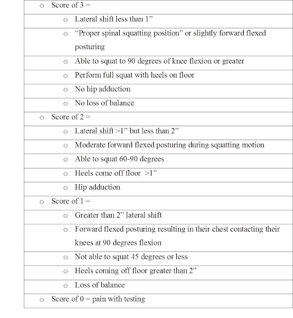
Scoring of the Movement Assessment (MA)

Before we begin the review of each of the tests, it is important to understand the scoring system for the MA.  The scoring system is important for the purposes of quantifying the movements themselves, but also for educating the patient or athlete about his or her movement patterns and finally, for establishing a baseline against which future measurements can be compared.  The MA is scored on a 0 to 3 scale using the criteria below, but keep in mind that these are not exhaustive lists for scores of 1 and 2.  Detailed scoring by deviation is listed for each individual test in the paragraphs that follow.


TEST #1:  Full Squat Test (FST) – the full squat test is used to assess how an athlete moves his or her body through a full squatting motion.  This test is crucial because it lets us know about the athlete’s balance, the flexibility of their hips, knees and ankles, the mobility of the spine and hip, and strength and endurance as they move the body’s center of gravity through a full > 90 degree squatting motion for multiple repetitions.  It also provides us with crucial information about exactly where the athlete’s movement is breaking down and whether that movement is breaking down in eccentric or concentric phases.  Using this information, we can identify not only what needs to be strengthened in the kinetic chain, but how it should be strengthened (eccentrically or concentrically).  During the full squat test, the athlete is asked to perform a full squat with little instruction about the depth of the squat expected, or the foot or knee placement.  The goal is to assess how a given subject will naturally perform the movement when given the opportunity.  The verbal instruction provided is “with your feet shoulder width apart, please perform a full squat.”  The athlete is viewed from both the anterior and posterior position.  They are asked to perform 10 repetitions facing toward and 10 repetitions facing away from the observer. 

During the full squat test, you are assessing the depth of the squat to 90 degrees, whether or not the heels remain on the floor, what the knee alignment is throughout the descent and then the ascent, and whether or not there is a lateral shift in the body at any time during the motion.  You are also assessing whether or not there is any change in mechanics when multiple repetitions are performed.

Clinical Implications of Full Squat Test

There are numerous deviations that show up in athletes performing squats.  Below is a list of some of the most common deviations associated with the Full Squat Test and the associated clinical implications.

Lateral deviation – or simply shifting the weight more to one side vs. the other.  This creates a tremendous amount of imbalance in the loading of the entire system and can contribute to significant strength deficits and a subsequent decrease in power output.  In the subject pictured, you will notice that she shifts her weight to the right side by ~3.5 inches when she squats.  This greatly increases the stress to the right side of the body and can result in significant muscular imbalances between the left and right sides of the body in strength, endurance and flexibility.

Some of the factors that can contribute to this shift include limited range of motion, decreased proprioception, pain, poor quadriceps strength and poor motor planning (or just bad training habits which lead to poor motor planning).  It is important to determine if the athlete is having pain with this or any action used to assess movement by simply asking “does this movement cause you pain?” 

Suggested Corrective Exercise:  When athletes present with this type of movement pattern, one of the most important things to consider doing to improve is the “Squat Neuromuscular Retraining Exercise” or “SNMR” (which will be described in later sections).  Along with that, it is important to incorporate dynamic stretches utilizing the principles of Squat Neuromuscular Retraining (SNMR), Lumbar Hip Disassociation, and the Single Leg with Dynamic Lower Extremity Movement exercise progression.

Increased trunk flexion – or excessive flexing of the lumbar spine which results in bending forward at the waist as opposed to movement from the hip.  This results in vertical displacement of the upper body or exaggerated knee flexion.  This deviation can come in many forms and can present in many different ways.  Two of the most common signs of this type of deviation are flexing forward in the lumbar spine instead of flexing at the hip and the chest contacting the knees at the end of the squat.  Increased trunk flexion directly impacts the lumbar spine through abnormal loading of the lower back.  Over time, this increases wear and tear on the lumbar spine and also significantly impacts lower extremity strength and power gains since the lower back bears load increases associated with additional training, instead of the hips, gluteal muscle groups, the quadriceps and hamstrings and the lower legs.  When athletes present with these movement patterns, there very often tends to be difficulty in differentiating lumbar movement from hip movement.  These athletes have a difficult time discerning the difference between motion that comes from the hip versus motion that comes from the lumbar spine.  In these cases, athletes can benefit from training in “lumbar hip disassociation”.  There also needs to be a determination if the lack of movement in the hips and/or knees is the result of decreased range of motion in the knee or hip, pain or tightness.  Simply asking “does this movement cause you pain,” and checking the range of motion in the knee and hip manually can provide insight into the reasons an athlete might perform a squat in this fashion. 

Suggested Corrective Exercise:  Training in lumbar hip disassociation as well as adding dynamic stretches (specifically the sumo squat), and the Single Leg with Dynamic Lower Extremity Movement exercise progression to the training plan will aid significantly.

Limited range of motion – or inability to perform the squatting motion throughout the full range without falling can be a result of limited flexibility of the quadriceps, hamstrings, calves, hip and lumbar spine as well as decreased strength gains and power output from the lower body.  Limitations in range of motion can result from pain, poor joint mobility of the hip, lumbar spine, foot and ankle or soft tissue limitations (i.e. decreased gastrocnemius or quadriceps flexibility) and poor balance.  Limitations in range of motion can show up in a lot of ways, whether it is in decreased depth of the squat (limited knee flexion) or loss of balance during the motion.  The subject in this picture has a decrease in knee flexion angles and due to the excessive weight shift forward, he lost his balance as well. 

Suggested Corrective Exercise:  When athletes present with these movement patterns, some treatment options to consider include squat neuromuscular retraining (SNMR), dynamic stretches, manual stretching and mobilization to the limited joints or tissue. 

Heels coming off floor – or inability to perform a full squat without the heels coming off of the floor can result in limited range of the squat which limits strength gains, flexibility and power output.  Heels coming off the floor can be caused by limited range of motion of the gastrocnemius or soleus/deep posterior compartment, poor balance or limited range of motion at the foot and ankle. 

In this picture, this subject has full knee flexion but lacks flexibility of the calf muscles which limits her from getting full range of motion and causes her heels to come off the floor during the squat.  When athletes present with this type of movement pattern, some treatment considerations include determining if the lack of motion originates at the knee or ankle. 

Suggested Corrective Exercise:  The addition of dynamic stretches, including manual stretching in the routine, stretching the gastrocnemius and soleus/deep posterior compartment and using the Single Leg with Dynamic Lower Extremity Movement exercise progression can be beneficial.

Hip adduction/femoral internal rotation – or the knees coming together with descent (on the way down) or ascent can have a direct impact on how forces are absorbed through the knee, hip and low back as well as have a negative impact on strength and power output. This movement pattern can have a huge impact on the integrity of the ligaments of the knee and can put the anterior cruciate ligament at risk.  Hip adduction and femoral internal rotation can be the result of poor core stability, decreased proprioception or decreased gluteus medius strength.

Suggested Corrective Exercise:  When athletes present with these movement patterns, there needs to be a component of hip (especially gluteus medius) and other core strengthening, and the Single Leg with Dynamic Lower Extremity Movement exercise progression.

Loss of balance – or the athlete is not able to go through a full range of motion without a loss of balance, often resulting in falling backwards or falling down.  Loss of balance has a significant impact on athletic performance and the ability to generate maximal force or power and also tells you a lot about an athlete’s sense of body awareness.  Loss of balance can result from lack of dynamic or static balance, lack of stability of the core or hip, lack of range of motion at the lumbar spine, hip, knee or foot/ankle or poor training technique. 

Suggested Corrective Exercise:  These athletes benefit greatly from the addition of the SNMR, lumbar hip disassociation training, core training, gluteus medius strengthening and dynamic stretches (specifically the sumo squat).

Dr. Nessler is a practicing physical therapist with over 17 years sports medicine clinical experience and a nationally recognized expert in the area of athletic movement assessment.  He is the developer of an athletic biomechanical analysis and author of a college textbook on this subject.  He serves as the National Director of Sports Medicine for Physiotherapy Associates, is a Safety Council Member for USA Cheer National Safety Council and associate editor of the International Journal of Athletic Therapy and Training. 

Monday, January 4, 2016

How to Identify Pathological Movement - Part II

Last week we discussed 3D technologies that are available to assess pathological movement in our athletes.  This week we will look at some 2D technologies as well as movement assessment specifically. 

Dartfish Video Technology

Most of us in the rehabilitation and sports performance setting are more familiar with 2D technologies.  Although there is a plethora of them on the market, one of the most recognized and most widely used in professional athletics is Dartfish.  In December of 1998 Dartfish was founded to develop a commercial product called SimulCam™ and other digital imaging applications for use in movement analysis and sports.  SimulCam™ technology was followed by StroMotion™ technology, which was launched in January 2001 and allowed viewing and movement analysis in much more depth.  Based on its technological know-how, Dartfish soon branched out into other growth areas such as interactive internet content enhancements and the development of cutting-edge sports training applications. Today, these technologies are widely recognized for exclusive televised broadcast footage of sporting events and breakthrough training applications for sports, education, healthcare and many other applications.

Using Dartfish technology affords users the ability to capture movement patterns and then review those movement patterns using both still pictures and video.  Dartfish also allows us to discretely, objectively measure some of the pathokinematic movement patterns that we see in the practical setting.  The ability to playback video as well as show still pictures of pathokinematics is a wonderful tool to use for patient and athlete education after measurements are made.  Since Dartfish can only provide one dimensional views of movement, accurate measurements of the components of rotation, however cannot be captured.  Although movements such as these can be visualized and described using the video captured with Dartfish software, actually quantifying these types of movements using numerical descriptors typically requires a three dimensional view, like those you can get with a Viacom multi-camera system. 

Dartfish video software comes in several versions designed to meet specific needs.  Fees involve purchase of a license and depending on the version of the program cost between $1200 and $4000.  In comparison to the biomechanics lab this is fairly inexpensive, but the cost can still be prohibitive for some users, such as high school athletic programs and the like.  In a clinical and performance setting though, it is an extremely valuable tool at a reasonable price which allows objective quantification of assessment findings, easy and effective patient and athlete education, and finally, a way to measure progress after application of targeted individualized treatment and athletic improvement plans. Its multi-functionality also allows it to be used indoors and outdoors but also has some limitations.

  • Time need for proficiency of use – it takes time to perfect and become efficient with this technology.
  • Angles – although it has the ability to measure angles, measurement of angles in 2D (using this or other technology) is just not accurate.  Inter and intrarater measurements have an error of 10-15 degrees.
  • Tracking – this is a 2D technology that tracks of pixels and unfortunately it is not accurate for tracking of movement.  For objects with a lot of contrast it is more accurate but in tracking limb movement it is not as accurate.
Despite the limitations, this is a great tool to not only assess movement but can also be used as a very effective tool in educating athletes regarding pathological movement.

Movement Assessment (MA)

Over the course of the last 15 years, we have assessing movement in order to capture and quantify pathokinematic movement patterns that we know lead to certain kinds of injury and limit athletic performance.  Movement assessment is a tool that can be used in conjunction with Dartfish video software, a laptop and a small digital video camera quickly and easily in the field under all kinds of conditions.  Relatively inexpensive, these tools can be used together in the clinical setting as well   For practical use in working with athletes to prevent injury, rehabilitate, or improve sports performance both on and off the field, movement assessment in conjunction with technology is an excellent tool to capture, quantify and then analyze movement patterns.  From there it is then possible to design and develop individualized training or rehabilitation protocols that address weaknesses in the kinetic chain that can only be seen when an athlete is in motion.

Any MA should consist of at least four essential movements that have a high correlation and carryover to sport and which allow us to assess pathokinematics.  A MA also provides us with a clearer indication of the exact location of the break down of normal movement patterns.  Using Dartfish video technology or 3D technology, we can video the athlete performing the MA to capture him or her in motion.  We can then use the video and/or freeze frame the movements in order to analyze both static positions and movement angles.  We can then assign measurements to both the static positions and the motion we capture on video.  Coaches, athletic trainers, physical therapists and others who work with the athlete can then use these measurements to formulate assumptions about the underlying structures of the body that facilitate or impede his or her motion. 

Finally, video footage and still pictures can be used to educate the athlete on the movement patterns he or she uses in sport and other activities, and how they might limit performance and lead to pain and/or injury.  This enables the coach, athletic trainer, or physical therapist and the athlete to begin dialogue within a common framework and understanding of where the athlete is today and where we want him or her to be in the future on these 6 essential movements.  A Corrective Exercise Program can then be designed and implemented cooperatively, (which we will discuss later), and progress can be measured periodically using the same tools.

A MA should consist of some of these foundational movements.  The Full Squat Test, the Single Leg Squat Test, the Plank Test and the Side Plank Test.  These should be done in a progressive sequence where the second test is hard than the first and the third harder than the second.  Completing each test in the progression requires greater levels of strength and allows us to make assumptions about how the body responds to increases in force demands and the impact fatigue has.  Considering fatigue, there should be minimal to no rest between each test.

Next week we will review each of the tests.  For each of the MA tests discussed here, we will review the test itself in detail, the deviations usually seen with the test, as well as the clinical implications of those deviations.  We will also briefly discuss a few of the treatment or training options associated with each test.  As noted earlier, the treatment/training options will be reviewed and discussed in further detail in future discussions.
Dr. Nessler is a practicing physical therapist with over 17 years sports medicine clinical experience and a nationally recognized expert in the area of athletic movement assessment.  He is the developer of an athletic biomechanical analysis and author of a college textbook on this subject.  He serves as the National Director of Sports Medicine for Physiotherapy Associates, is a Safety Council Member for USA Cheer National Safety Council and associate editor of the International Journal of Athletic Therapy and Training.